DEGREES OF COMPARISON OF ADJECTIVES In English, as in Russian, ...


The femur is one of the strongest in the body, so its fracture can occur only with serious injuries or pathologies. Main symptoms: pain, inability to stand on the injured leg. A hip fracture in a dog needs surgical treatment.
The femur is long tubular, the upper end has a spherical head and is involved in the formation of a multiaxial hip joint, reinforced by an intracapsular round ligament.
The lower end of the thigh has two rounded blocks to form the femoral and tibial joints.
The upper end of the thigh, in addition to the head with a hole under it (for attaching the round ligament), has a neck, large and small skewers for attaching the gluteal muscle group. Therefore, in Perthes disease, a fracture of the femoral neck is more often observed, as places with the greatest load. The trochanters are connected by an intertrochanteric ridge, which creates the boundaries of the trochanteric fossa.
The lower end of the femur has two ridges of the same size for the patella, between which there is a groove for it to slide during movement. The lower end ends with two condyles - rounded elevations for attaching the tibia. Between them is the intercondylar fossa.
According to the etiology, fractures can be divided into two large groups: traumatic and pathological.
The causes of traumatic fractures are varied:
Several diseases lead to pathological fractures:
In addition to classification for reasons, there is a division according to the type of fracture:
The most common symptom is severe pain. She does not allow the dog to lean on the limb. When trying to feel the injured leg, the dog behaves aggressively, it may even bite the owner. When examining a limb, one can see edema, hematoma, and in severe cases, a wound, sometimes with a bone fragment in it. Often, the asymmetry of the hind limb is immediately visible, but sometimes such symptoms are not detected.
Since the dog can behave aggressively, you should immediately put a muzzle on it so that it does not bite the owner or the doctor. The first aid is to fix the limb. To do this, you need to take a long, strong and not flexible stick and tie it to the dog. On the one hand - to the knee, on the other - to the pelvis in the region of the hip joint.
You can not try to set the bone yourself, it will cause an incredibly strong pain reaction.
In addition, you can damage blood vessels and nerves with fragments of bones. The bone should be set by a specialist and only after an X-ray. X-ray is done in two perpendicular planes.
After fixing the limb, you need to deliver the dog to the veterinarian as soon as possible. Large pets are carried on a piece of plywood, a stretched blanket, and other makeshift stretchers. It is not recommended to do anesthesia, as this can lead to incompatibility of the drugs that the doctor will use.
Treatment at home is impossible, since the condition with fractures is too dangerous. First of all, anesthesia and general anesthesia are carried out, so that the dog allows itself to be examined calmly. Next, an x-ray is taken to determine the position of the bone fragments. Often, in case of fractures, surgery is done immediately.
With incomplete fractures (cracks) or with closed fractures without displacement of the bones, plaster is applied or a splint is created. However, this method is not always effective, since the dog may try to remove the cast, thereby injuring his limb again. In this regard, veterinarians prefer to perform operations.
The operation is done under general anesthesia, all methods of fixation can be divided into two large groups.
The first incision is made at the site of the fracture. Through it, small fragments of bones, blood clots are removed, with an open fracture, they are treated with antibiotics. Next, the upper and lower bone fragments are removed into the wound so that it is convenient to manipulate them.
Then, using a stylet and a drill, a hole is made through the bone fragment near the upper head of the femur. It is made from the inside, through the medullary canal, through the first incision, this is necessary to introduce a conductor - a metal wire that will set the direction of the pin.
The conductor is pushed into the bone through the fracture site to the very top, so that it comes out from the upper end of the bone in the zone of the vertical cavity (between the head of the bone and the greater and lesser trochanters). When the conductor rests against the skin of the buttock, a second incision is made in this place.
A pin is inserted through the second incision, with the help of a conductor it is given a direction. If necessary, help the pin with a hammer to enter the bone. It is pushed until it comes out of the upper fragment by half a centimeter. After that, the ends of the bones are connected and the pin is inserted completely.
If in the first case the pin is inserted into the bone, then according to this method, the fixator is attached from above to the bone. This method is preferred for small breeds, as their bones are thin and do not allow the use intraosseous method. The latch itself can be in the form of a plate, beam or rings.
An incision is made at the fracture site, then the injured area is cleaned of small bone fragments, crushed tissues, blood clots. The retainer is attached using screws, rings, spokes and other devices. It all depends on the type of fixing device.
Further care includes rest, restriction of movement for the first week. The dog itself will determine the time when you can lean on the paw. On the third day, a callus is formed between the fragments of bones, consisting of cartilage tissue, which begins to ossify after 8-10 days. Complete fusion of the bone occurs in 25-45 days.
(treatment of non-union of bone fragments of the bones of the forearm).
If we summarize the complications in the treatment of fractures of the bones of the forearm in dogs of dwarf breeds, then we can state that the underlying cause is the “human factor”: unstable fixation of bone fragments among themselves, impaired vascularization of fragments, thermal burn of the bone, use of large implants ( Yagnikov S.A., Kozhushko P.S. Complications in dogs of dwarf breeds in the treatment of fractures of the bones of the forearm. Russian veterinary journal. Small pets. No. 1. 2014 pp. 6 – 10. )
The VetProfAlliance Center for Veterinary Surgery has accumulated extensive experience in the treatment of complicated fractures (nonunions) of the forearm bones in dwarf breed dogs (see pictures).
Rice. 1. Vascular hypertrophic non-union of the bones of the forearm. Fixation with a double-sided single-plane clamp. Bone grafting. Fragment consolidation.
Rice. 2. Vascular oligotrophic nonunion after osteosynthesis with a Kirschner wire. Combined osteosynthesis with Kirschner wires and a two-sided single-plane fixator with bone grafting. Fragment consolidation.

Rice. 3. Avascular, dystrophic nonunion after plate osteosynthesis. Removal of implants. Extrafocal osteosynthesis with a bilateral single-plane fixator with bone autoplasty. Consolidation of fragments of the radius and ulna.

Rice. 4. Vascular, dystrophic non-union of forearm bone fragments after intramedullary osteosynthesis with a Kirschner wire. Avascular, atrophic nonunion of the radius. Removing the Kirschner wire. Extrafocal osteosynthesis with a bilateral single-plane fixator, with bone grafting with spongy autologous bone. Consolidation of fragments of the radius.

Rice. 5. Avascular, dystrophic non-union of the bones of the forearm in a dwarf breed dog after intramedullary osteosynthesis with a Kirschner wire. Extrafocal osteosynthesis with a two-sided single-plane fixator with bone autoplasty. Fracture consolidation.

Rice. 6. Inadequate osteosynthesis with a Kirchner wire. Combined osteosynthesis with a Kirschner wire and a plate for 2.0 mm screws. Fracture consolidation.

Rice. 7. Inadequate osteosynthesis with a Kirschner wire. Limb deformity. Combined osteosynthesis with a Kirschner wire and a plate for 2.0 mm screws. Fracture consolidation.

Rice. 8. Bone osteosynthesis for fracture of the forearm in dogs of dwarf breeds. Fragment consolidation.
Conclusion:
For the treatment of fractures, the use of an immobilizing dressing (gypsum) has traditionally been used, this method of treatment has a number of disadvantages - the development of atrophy of the muscles of the limb, frequent malunion of bones, the formation of bedsores under the bandage, impaired blood supply to bones and soft tissues. All these complications led to the abandonment of the widespread use of gypsum for the treatment of fractures, so now this treatment method is used only for the treatment of cracks. A more modern method of treating fractures is osteosynthesis- surgery for surgical comparison of bone fragments with the use of fixing metal structures.
Types of osteosynthesis:
1. Intramedullary osteosynthesis - used to treat fractures of long bones. With this method, a special pin or needle is installed inside the bone. But there are limitations to this method - for example, it is not suitable for the treatment of fractures of the pelvis, skull, spine, jaw, as well as for the treatment of comminuted fractures.
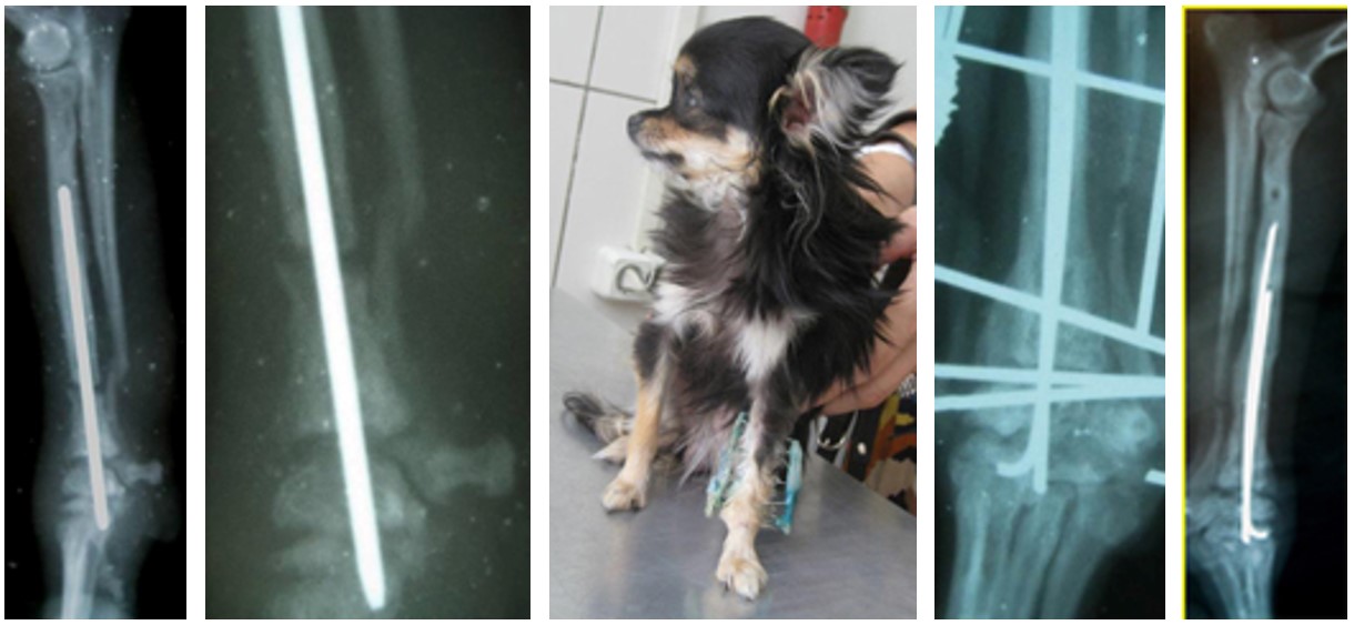
ferret hip fracture

The use of intramedullary osteosynthesis for hip fracture
2. Bone osteosynthesis - with this method, a metal plate is attached to the bones with the help of special bolts. As a result, good stabilization of bone fragments is achieved. This method can treat not only fractures of tubular bones, but also injuries of the pelvis, skull, spine, scapula, etc. The negative side of this method is the rather high cost of the operation associated with the use of expensive materials (plates, bolts and special tools).

Fracture of the forearm in a dog

Bone osteosynthesis

Gunshot wound to the lower jaw with a fracture of both branches of the lower jaw

View after osteosynthesis
3. Extrafocal osteosynthesis - is used to treat not only fractures, but also dislocations, and consists in passing the spokes through the bone above and below the fracture site with their subsequent fixation from the outside with a special polymer. The advantages of this method are the relative cheapness of consumables, the speed of the operation, and the reliability of fixing debris. The disadvantage of this method is the impossibility of applying an external fixation device in large and giant breeds of dogs.

X-ray after extrafocal osteosynthesis
4. Combined osteosynthesis - consists in the use of several of the above methods and is used mainly for complex comminuted fractures.

A cat with a compound comminuted fracture of the femur

Cat after combined osteosynthesis

Intercondylar fracture of the humerus in a dog

After osteosynthesis
Separately, it is worth considering fractures of the pelvis. As a rule, such injuries are received by dogs as a result of car accidents, and cats by falling from a great height. In case of damage to the pelvic bones, fractures are usually multiple, which makes them the most difficult in the practice of a traumatologist.

Multiple pelvic fractures in a dog. On the right - a fracture of the pubic and ischial bones, on the left - a fracture of the acetabulum.

The same dog after osteosynthesis

Use of a compression plate for a complex fracture of the acetabulum
Our veterinary clinic has accumulated extensive experience in the use of all types of osteosynthesis in animals of all sizes, which allows us to approach the treatment of each case individually and recommend the most optimal method of reconstructive surgery.
Prices, rub.
The price does not include consumables and additional work
Question answerIs it possible to fix an old fracture (the radius of the front right paw in a dog)? If so, what is the name of this operation? A week later, we were booked in for an examination and an x-ray of an old fracture, we are waiting for what they say. But I would also like to get an answer to the question above ... The fracture has grown together crookedly, the dog is from the street. Julia
Q: Is it possible to fix an old fracture in a dog?
Hello! Maybe. This is metal osteosynthesis. But the only way to tell for sure is from the picture.
Hello. Tell me the approximate amount of total expenses, including additional ones, for prosthetics of the cat's paw. Amputated as a result of falling into a trap, in the area of the wrist.
Question: Can you tell me the approximate amount for prosthetics for a cat's paw?
Hello! For prosthetics please email us. [email protected] with a note to Sergey Sergeevich Gorshkov. It needs to be reviewed and reviewed. On the offhand, no one will say the approximate cost.
S.A.Erofeev., N.V.Petrovskaya, A.A.Emanov acad. G.A. Ilizarov, Kurgan
Source: materials of the Moscow International Veterinary Congress
Treatment of animals with fractures of both bones of the forearm remains an urgent problem of modern veterinary traumatology. With this type of injury, the musculoskeletal function of the thoracic limb as a whole is excluded, and without the provision of qualified veterinary care, the animal may remain "disabled". According to our data, fractures of the bones of the forearm account for 15% of the total number of fractures of long tubular bones. For the treatment of fractures of the forearm, we use the method of transosseous osteosynthesis, developed by G.A. Ilizarov in the middle of the last century. This method allows to provide equally favorable conditions for the fusion of fragments of the radius and ulna. One of the main conditions for the successful use of transosseous osteosynthesis is knowledge of the topographic anatomy of the segment. This is necessary in order to exclude damage to blood vessels and nerves during the wires, avoid trauma to the main muscle masses and prevent fixation of the tendon-aponeurotic formations.
The aim of this study was to develop a technique for conducting wires at different levels of the forearm, and to analyze the results of treatment of dogs with fractures in this area.
The proposed safe technique for osteosynthesis of the forearm is based on the literature data on anatomy, topographic anatomy (A.I. Akaevsky., 1984, B.M. Khromov et al., 1972, Coy Alpha, 1996, J.S. Boyd., 1998, H.A. Slesarenko, 2000 ), as well as on the results of our own studies of angiograms, anatomical preparations and Pirogov sections.
At the proximal level of the forearm, fragments are fixed with a pair of mutually intersecting wires, at an angle of 65-70˚, which, in order to avoid getting into the elbow joint, are located 1.0-1.5 cm distal to the head of the radius. In the absence of the possibility of palpation of the head of the radial bone, bone protrusions serve as reference points for the wires: the epicondyles of the humerus and the olecranon. The needles must be passed through the middle of the diameter of the bones at a distance from each other equal to the thickness of the support. We fix the spokes in a taut state on different planes of the arc, which faces the open part cranially, in order to preserve the anatomical and physiological function of the elbow joint.
We pass one of the spokes through both bones of the forearm from the lateral surface, in the oblique-sagittal plane. The injection point is located at the level of the radial roughness, from which we retreat to the middle of the radius, in the caudo-medial direction. On its way, the pin passes through the common extensor of the fingers, superficial and deep flexors of the fingers, it is necessary to bypass the common interosseous artery.
The second needle - through the ulna from the medial surface, in the frontal plane. We make an injection in the middle of the radial flexor of the wrist, leaving the neurovascular bundle (median artery, vein and nerve) 2-3 mm more cranially. On its way, the spoke inevitably fixes the radial and ulnar flexors of the wrist.
At the distal level of the forearm, three wires are passed through the fragments, which we fix in the annular support. Guidelines for their implementation are the tops of the styloid processes of the radius and ulna, from which we retreat 2-2.5 cm. In young animals, the crosses of the spokes must be carried out outside the growth zones. One needle is passed through both bones in a plane close to the frontal, with an injection point on the side of the ulna. In this case, the pin passes through the distal intertendinosus muscle, bypassing the cranial interosseous and ulnar arteries.
The other two spokes - through the radius and ulna in the oblique-sagittal planes, with a decussation angle of 65-70˚ relative to each other, while the medio-caudal surface remains intact, on which the tendons of the flexor muscles are grouped.
The plane of the wires at different levels of the segment should be perpendicular to the axis of the fragments.
Regardless of the level of the fracture, through the diaphyseal section of each fragment, to complete their reposition and ensure the stability of fixation, we carry out the spokes with subsequent fixation in the corresponding ring supports. The plane of their conduction, as a rule, is frontal, retreating 1.5-2 cm from the fracture line. The projection lines of the median artery and nerve serve as a guideline for determining the injection points. Depending on the clinical situation, for reposition and fixation of fragments, we use spokes with a thrust platform.
The age of the animals ranged from 3.5 months to 7 years. The causes of fractures in 57.9% of cases were domestic trauma, in 34.2% - road and 7.9% - gunshot wounds. The dogs with closed injuries were the most frequent (81.6%). According to the nature of the fractures, they were distributed as follows: transverse - 51.3%; oblique - 16.2%; comminuted - 18.9%. Damage to the lower third of the forearm accounted for 63.1% of cases, the middle third - 23.7% and the upper third - 13.2%. According to Coy Alfa (1996), the predominance of lesions in the lower third of the segment is due to the absence of muscle bellies in the distal forearm.
The reception of the animal began with the collection of anamnesis, general examination, assessment of the local status and radiographic examination of the damaged segment. Contraindications to osteosynthesis were damage to internal organs, concomitant infectious and skin diseases. Surgical intervention was carried out on average on the third day after the injury with the obligatory written consent of the owner.
Based on the X-ray picture of the damaged segment, in two standard projections, before the operation, depending on the level of the fracture, the apparatus module was assembled. In case of damage to the middle third of the forearm, the apparatus consisted of four supports (arc, 3 rings); for fractures of the upper and lower thirds - from three supports (arc, 2 rings). Depending on the length of the forearm, in small dogs, two supports (arc, ring) and brackets were used.
Transosseous osteosynthesis was performed in the operating room in compliance with the rules of asepsis and antisepsis. Under intravenous barbituric anesthesia with preliminary premedication (rometar, droperidol, atropine) in appropriate doses. The animal was fixed in a lateral position on the side of a healthy limb. The operating field was treated with 5% alcohol solution of iodine.
Focusing on the data of the X-ray picture, a preliminary manual reposition of the fragments was performed, which made it possible to eliminate the gross displacement. The application of the apparatus began with passing one wire through the ulna at the proximal level, and through both bones at the distal level. The apparatus was centered relative to the axis of the forearm and distraction was carried out along the rods to eliminate the displacement along the length. Then, at the proximal level, a pin was passed through both bones, and at the distal level, two pins were passed separately through the radius and ulna. In case of damage to the epiphyses of the bones of the forearm, a pin was passed through the metacarpal bones for stable fixation of the distal fragments. The quality of reposition was controlled by palpation and using injection needles. At 1-1.5 cm on both sides of the fracture line, one needle was injected perpendicular to the axis of each fragment. The location of the needle cannulas at the same level indicated the absence of fragment displacement.
This technique excluded radiological control during the operation. Further conduction of repositioning-fixation pins in the middle third depended on the displacement of the fragments. In each support, the spokes were tensioned using a calibrated spoke tensioner. The load on the spoke depended on the type (arc, ring), diameter, thickness and material from which the support was made. So, for example, with a diameter of 100 mm, a thickness of 5 mm in a steel arc, the first spoke was tensioned with a force of 90 kg, the second - 80 kg, in the ring - the first - 100 kg, the second - 110 kg. The operation ended with a control X-ray of the segment.
Further treatment of animals was carried out on an outpatient basis. Owners received advice on dog care:
In the first two days, painkillers (analgin, baralgin);
- with open fractures - a course of antibiotic therapy;
-toilet of soft tissues near the spokes;
- control over the state of the device and its isolation with a cloth cover;
- wiring on a leash and passive work with the joints of the chest limb;
Medical control, if possible, was carried out weekly, X-ray - one week after the operation and every two to three weeks of subsequent fixation. Fracture consolidation was determined by a combination of radiological and clinical signs of union. The duration of fixation of the limb in the apparatus before the onset of fusion ranged from 14 to 65 days, which depended on the age of the animal and the severity of the injury. The average time for union was 32.8±1.7 days.
Thus, the use of transosseous osteosynthesis for fractures of the forearm bones in dogs provides reposition, stable fixation of fragments and preservation of the blood supply to the segment. The method allows to quickly restore the function of the injured limb and achieve positive anatomical and functional results of treatment.
Summary
Yerofeyev S.A., Petrovskaya N.V., Yemanov A.A. Transosseous osteosynthesis of canine forearm eones. Russian Ilizarov Scientific Center "Restorative Traumatology and Orthopaedics", Kurgan, Russia.
Thus, use of transosseous osteosynthesis for fractures of canine forearm bones provides reposition, stable fixation of fragments and maintenance of segmental blood supply. The technique allows to restore function of the limb involved and obtain positive anatomic-and-functional results of treatment in a short time.
Fracture of the distal part of the ulna and ulna is the most common in our practice, fractures in dogs of dwarf breeds such as Yorkshire Terrier, Toy Terrier, Spitz, etc.
Such fractures occur when animals fall from a height, sometimes for a fracture it is enough to land unsuccessfully from a sofa or chair.
In the event of a fracture, the owner of the animal must fix the limb to prevent injury to soft tissues by bone fragments.
The most effective way to treat such fractures in dogs of dwarf breeds is osteosynthesis using a transosseous, external fixator.
Similar fractures in toy breed dogs are diagnosed based on clinical signs such as pain, swelling, shortening of the paw, and the dog completely stops using the limb.
For the final diagnosis and development of tactics of the operation, it is necessary to conduct an X-ray examination.
In most cases, we are faced with a fracture of the distal third of the ulna and radius with displacement, often sharp bone fragments perforate the skin.
Intermedullary osteosynthesis. With this type of treatment, a pin is inserted into the intermedullary canal of the bone. Due to the fact that one hundred diameter of the ulna and radius
Dogs weighing 1.5-2 kg are only 2-3 mm, so the introduction of an intraosseous fixator is extremely difficult, as when the fixators are introduced, the blood supply to the bone tissue is disrupted, which can subsequently lead to delayed bone fusion in dogs or even to bone lysis.
Bone osteosynthesis with a plate. This method is the gold standard for the treatment of such fractures in large dogs, but in toy breeds this method is also difficult due to the small diameter of the bone.
Bandages, splints. It is not recommended to use such methods. with these methods of treatment, an exact reposition (comparison) and immobilization (fixed fixation) of inert fragments are not achieved. Correct fusion usually does not occur.
Extrafocal osteosynthesis with a rod device. This method allows you to securely fix bone fragments with minimal trauma to the joints and soft tissues, as well as minimizing trauma to the bone itself, and the functionality of the limb is not disturbed.
The surgical operation consists of the following steps:
In the postoperative period, prophylactic administration of antibiotics and the necessary analgesia are necessary.
Removal of the device is made for 30-45 days. The dog begins to use the limb already for 4-5 days.
In all cases of treatment, good treatment results were achieved.
Complications include early loosening of the pins, breakage of the fixator, repeated fractures or fractures of the bones at the site of the pin.
The disadvantages of the method are the laboriousness of the operation, the multi-stage treatment and the need for postoperative care for the device.
Extrafocal osteosynthesis with rod devices in the treatment of forearm fractures in miniature dogs is a low-traumatic, reliable method that gives good functional results.
DEGREES OF COMPARISON OF ADJECTIVES In English, as in Russian, ...

In the early days of comics, there were several female wrestlers with...

The message about Beethoven, summarized in this article, will tell you about the great ...