In order to diversify the festive table, a woman often tries...


What are mitochondria? If the answer to this question is difficult for you, then our article is just for you. We will consider the structural features of these organelles in relation to the functions they perform.
But first, let's remember what organelles are. This is what permanent cellular structures are called. Mitochondria, ribosomes, plastids, lysosomes... All these are organelles. Like the cell itself, each such structure has a general structural plan. Organelles consist of a surface apparatus and internal contents - the matrix. Each of them can be compared to the organs of living beings. Organelles also have their own characteristic features that determine their biological role.
Organelles are divided into groups based on the structure of their surface apparatus. There are single-, double- and non-membrane permanent cellular structures. The first group includes lysosomes, the Golgi complex, the endoplasmic reticulum, peroxisomes and various types of vacuoles. The nucleus, mitochondria and plastids are double-membrane. And ribosomes, the cell center and organelles of movement are completely devoid of surface apparatus.
What are mitochondria? For evolutionary teaching, these are not just cell structures. According to the symbiotic theory, mitochondria and chloroplasts are the result of metamorphoses of prokaryotes. It is possible that mitochondria originated from aerobic bacteria, and plastids from photosynthetic bacteria. Proof of this theory is the fact that these structures have their own genetic apparatus, represented by a circular DNA molecule, a double membrane and ribosomes. There is also an assumption that animal eukaryotic cells subsequently evolved from mitochondria, and plant cells from chloroplasts.

Mitochondria are an integral part of the cells of the majority of plants, animals and fungi. They are absent only in anaerobic unicellular eukaryotes living in an oxygen-free environment.
The structure and biological role of mitochondria have long remained a mystery. They were first seen using a microscope by Rudolf Kölliker in 1850. In the muscle cells, the scientist discovered numerous granules that looked like fluff in the light. Understanding the role of these amazing structures was made possible thanks to the invention of University of Pennsylvania professor Britton Chance. He designed a device that allowed him to see through organelles. This is how the structure was determined and the role of mitochondria in providing energy to cells and the body as a whole was proven.

Let's consider what mitochondria are from the point of view of their structural features. These are double-membrane organelles. Moreover, the outer one is smooth, and the inner one has outgrowths. The mitochondrial matrix is represented by various enzymes, ribosomes, monomers of organic substances, ions and clusters of circular DNA molecules. This composition makes it possible for the most important chemical reactions to occur: the tricarboxylic acid cycle, urea, and oxidative phosphorylation.

Mitochondria membranes are not identical in structure. The closed outer one is smooth. It is formed by a bilayer of lipids with fragments of protein molecules. Its total thickness is 7 nm. This structure performs the functions of delimitation from the cytoplasm, as well as the relationship of the organelle with the environment. The latter is possible due to the presence of the porin protein, which forms the channels. Molecules move along them through active and passive transport.
The chemical basis of the inner membrane is proteins. It forms numerous folds inside the organoid - cristae. These structures significantly increase the active surface of the organelle. The main feature of the structure of the inner membrane is complete impermeability to protons. It does not form channels for the penetration of ions from the outside. In some places the outer and inner contact. A special receptor protein is located here. This is a kind of conductor. With its help, mitochondrial proteins, which are encoded in the nucleus, penetrate into the organelle. Between the membranes there is a space up to 20 nm thick. It contains various types of proteins, which are essential components of the respiratory chain.

The structure of the mitochondrion is directly related to the functions it performs. The main one is the synthesis of adenosine triphosphate (ATP). This is a macromolecule that is the main carrier of energy in the cell. It consists of the nitrogenous base adenine, the monosaccharide ribose and three phosphoric acid residues. It is between the last elements that the main amount of energy is contained. When one of them ruptures, a maximum of 60 kJ can be released. In total, a prokaryotic cell contains 1 billion ATP molecules. These structures are constantly in operation: the existence of each of them in an unchanged form does not last more than one minute. ATP molecules are constantly synthesized and broken down, providing the body with energy at the moment it is needed.
For this reason, mitochondria are called "energy stations." It is in them that the oxidation of organic substances occurs under the action of enzymes. The energy that is generated in this case is stored and stored in the form of ATP. For example, when 1 g of carbohydrates is oxidized, 36 macromolecules of this substance are formed.
The structure of mitochondria allows them to perform another function. Due to their semi-autonomy, they are an additional carrier of hereditary information. Scientists have found that the DNA of the organelles themselves cannot function independently. The fact is that they do not contain all the proteins necessary for their work, so they borrow them from the hereditary material of the nuclear apparatus.
So, in our article we looked at what mitochondria are. These are double-membrane cellular structures, in the matrix of which a number of complex chemical processes take place. The result of the work of mitochondria is the synthesis of ATP, a compound that provides the body with the necessary amount of energy.
Special structures - mitochondria - play an important role in the life of each cell. The structure of mitochondria allows the organelle to operate in a semi-autonomous mode.
Mitochondria were discovered in 1850. However, it became possible to understand the structure and functional purpose of mitochondria only in 1948.
Due to their rather large size, the organelles are clearly visible in a light microscope. The maximum length is 10 microns, the diameter does not exceed 1 micron.
Mitochondria are present in all eukaryotic cells. These are double-membrane organelles, usually bean-shaped. Mitochondria are also found in spherical, filamentous, and spiral shapes.
The number of mitochondria can vary significantly. For example, there are about a thousand of them in liver cells, and 300 thousand in oocytes. Plant cells contain fewer mitochondria than animal cells.
TOP 4 articleswho are reading along with this

Rice. 1. The location of mitochondria in the cell.
Mitochondria are plastic. They change shape and move to the active centers of the cell. Typically, there are more mitochondria in those cells and parts of the cytoplasm where the need for ATP is higher.
Each mitochondrion is separated from the cytoplasm by two membranes. The outer membrane is smooth. The structure of the inner membrane is more complex. It forms numerous folds - cristae, which increase the functional surface. Between the two membranes there is a space of 10-20 nm filled with enzymes. Inside the organelle there is a matrix - a gel-like substance.

Rice. 2. Internal structure of mitochondria.
The table “Structure and functions of mitochondria” describes in detail the components of the organelle.
|
Compound |
Description |
Functions |
|
Outer membrane |
Consists of lipids. Contains a large amount of porin protein, which forms hydrophilic tubules. The entire outer membrane is permeated with pores through which molecules of substances enter the mitochondria. Also contains enzymes involved in lipid synthesis |
Protects the organelle, promotes the transport of substances |
|
They are located perpendicular to the mitochondrial axis. They may look like plates or tubes. The number of cristae varies depending on the cell type. There are three times more of them in heart cells than in liver cells. Contains phospholipids and proteins of three types: Catalyzing - participate in oxidative processes; Enzymatic - participate in the formation of ATP; Transport - transport molecules from the matrix out and back |
Carries out the second stage of breathing using the respiratory chain. Hydrogen oxidation occurs, producing 36 molecules of ATP and water |
|
|
Consists of a mixture of enzymes, fatty acids, proteins, RNA, mitochondrial ribosomes. This is where mitochondria's own DNA is located. |
Carries out the first stage of respiration - the Krebs cycle, as a result of which 2 ATP molecules are formed |
The main function of mitochondria is the generation of cell energy in the form of ATP molecules due to the reaction of oxidative phosphorylation - cellular respiration.
In addition to mitochondria, plant cells contain additional semi-autonomous organelles - plastids.
Depending on the functional purpose, three types of plastids are distinguished:

Rice. 3. Plastids.
We examined the structural features of mitochondria - double-membrane organelles that carry out cellular respiration. The outer membrane consists of proteins and lipids and transports substances. The inner membrane forms folds - cristae, on which hydrogen oxidation occurs. The cristae are surrounded by a matrix - a gel-like substance in which some of the reactions of cellular respiration take place. The matrix contains mitochondrial DNA and RNA.
Average rating: 4.4. Total ratings received: 105.
Mitochondria are microscopic membrane-bound organelles that provide the cell with energy. Therefore, they are called energy stations (battery) of cells.
Mitochondria are absent in the cells of simple organisms, bacteria, and entamoeba, which live without the use of oxygen. Some green algae, trypanosomes contain one large mitochondrion, and the cells of the heart muscle and brain have from 100 to 1000 of these organelles.
Mitochondria are double-membrane organelles; they have outer and inner membranes, an intermembrane space between them, and a matrix.
Outer membrane. It is smooth, has no folds, and separates the internal contents from the cytoplasm. Its width is 7 nm, it contains lipids and proteins. An important role is played by porin, a protein that forms channels in the outer membrane. They provide ion and molecular exchange.
Intermembrane space. The size of the intermembrane space is about 20 nm. The substance filling it is similar in composition to the cytoplasm, with the exception of large molecules that can penetrate here only through active transport.

Inner membrane. It is built mainly from protein, only a third is allocated to lipid substances. A large number of proteins are transport proteins, since the inner membrane lacks freely passable pores. It forms many outgrowths - cristae, which look like flattened ridges. Oxidation of organic compounds to CO 2 in mitochondria occurs on the membranes of the cristae. This process is oxygen-dependent and is carried out under the action of ATP synthetase. The released energy is stored in the form of ATP molecules and is used as needed.
Matrix– the internal environment of mitochondria has a granular, homogeneous structure. In an electron microscope, you can see granules and filaments in balls that lie freely between the cristae. The matrix contains a semi-autonomous protein synthesis system - DNA, all types of RNA, and ribosomes are located here. But still, most of the proteins are supplied from the nucleus, which is why mitochondria are called semi-autonomous organelles.
Hondriom is a group of mitochondria that are concentrated in one cell. They are located differently in the cytoplasm, which depends on the specialization of the cells. Placement in the cytoplasm also depends on the surrounding organelles and inclusions. In plant cells they occupy the periphery, since the mitochondria are pushed towards the membrane by the central vacuole. In renal epithelial cells, the membrane forms protrusions, between which there are mitochondria.
In stem cells, where energy is used equally by all organelles, mitochondria are randomly distributed. In specialized cells, they are mainly concentrated in areas of greatest energy consumption. For example, in striated muscles they are located near the myofibrils. In spermatozoa, they spirally cover the axis of the flagellum, since a lot of energy is needed to set it in motion and move the sperm. Protozoans that move using cilia also contain large numbers of mitochondria at their base.
Division. Mitochondria are capable of independent reproduction, having their own genome. Organelles are divided by constrictions or septa. The formation of new mitochondria in different cells differs in frequency; for example, in liver tissue they are replaced every 10 days.
Molecular biology is the science that studies the role of mitochondria in metabolism. They also convert pyruvate into acetyl-coenzyme A and beta-oxidation of fatty acids.
| Table: structure and functions of mitochondria (briefly) | ||
|---|---|---|
| Structural elements | Structure | Functions |
| Outer membrane | Smooth shell, made of lipids and proteins | Separates the internal contents from the cytoplasm |
| Intermembrane space | There are hydrogen ions, proteins, micromolecules | Creates a proton gradient |
| Inner membrane | Forms protrusions - cristae, contains protein transport systems | Transfer of macromolecules, maintenance of proton gradient |
| Matrix | Location of Krebs cycle enzymes, DNA, RNA, ribosomes | Aerobic oxidation with the release of energy, the conversion of pyruvate to acetyl coenzyme A. |
| Ribosomes | Combined two subunits | Protein synthesis |
The common properties of mitochondria and chloroplasts are primarily due to the presence of a double membrane.
Signs of similarity also include the ability to independently synthesize protein. These organelles have their own DNA, RNA, and ribosomes.
Both mitochondria and chloroplasts can divide by constriction.
They are also united by the ability to produce energy; mitochondria are more specialized in this function, but chloroplasts also produce ATP molecules during photosynthetic processes. Thus, plant cells have fewer mitochondria than animal cells, because chloroplasts partially perform the functions for them.
Let us briefly describe the similarities and differences:
These organelles differ in their functions: mitochondria are intended for energy synthesis, cellular respiration occurs here, chloroplasts are needed by plant cells for photosynthesis.
The internal organization of an animal and plant cell can be compared to a commune, where everyone is equal and everyone plays one, very specific role, creating a balanced ensemble. And only one structure, the mitochondrion, can boast of a multiplicity of intracellular functions that determine its uniqueness and isolation, bordering on some self-sufficiency.
This structure was discovered in the mid-19th century, and for 150 years almost everyone believed that its sole function was to be the energy engine of the cell. Roughly speaking, the body receives nutrients, which, after a certain degradation, reach the mitochondria and then oxidative degradation of nutrients occurs, coupled with the storage of energy in the form of an energy-rich phosphorus bond in the ATP molecule. The body uses ATP energy everywhere, spending it on the conduction of a nerve signal, muscle contraction, heat generation, synthesis of necessary cellular components, destruction of unnecessary substances, etc. ATP is generated in the human body per day, weighing equal to the weight of the person himself, and this is mainly due to mitochondria . There is still debate about whether eukaryotic (nucleated) cells without mitochondria exist. While there is no clearly proven evidence of this, it is believed that nuclear cells without mitochondria do not exist.
There is still debate about whether eukaryotic (nucleated) cells without mitochondria exist. While there is no clearly proven evidence of this, it is believed that nuclear cells without mitochondria do not exist
The postulate of the dominant energy function of mitochondria in the cell somehow left in the shadows the long-proposed and universally supported theory of the bacterial origin of mitochondria. In a simple interpretation, it looks like this: about 600 million years ago in the so-called cell. heterotrophs, a bacterium is introduced that can utilize oxygen. There is a point of view that the appearance of a new type of bacteria inside a cell was caused by a constant increase in oxygen in the Earth’s atmosphere, which began to flow from the world’s oceans into the atmosphere about 2.4 billion years ago. The high oxidative capacity of oxygen posed a danger to intracellular organic and inorganic elements, and bacteria appeared that destroyed oxygen in the presence of hydrogen ions to form water. Thus, the oxygen content inside the cell decreases, and with it the likelihood of unwanted oxidation of cellular components decreases, which is probably beneficial for the cell.
The entry of bacteria into the intracellular niche also provided protection from external enemies (and the main enemies for bacteria are viruses, that is, phages). At the same time, it was allowed to release signaling protective substances into a limited intracellular volume; when bacteria existed in the “ocean”, the release of such signaling substances was irrational - they were immediately diluted in it. The life of intracellular bacteria in this niche provided certain advantages: the bacteria produce energy and organize a protein in their membrane that releases synthesized ATP into the cell’s cytoplasm, which the cell uses. As a result, there seems to be a balance: the cell gives the mitochondria nutrient substrates, the mitochondria gives the cell energy, which strengthens the theory of the symbiotic relationship between bacteria (they already become mitochondria) with the rest of the cell. The main arguments supporting the bacterial origin of mitochondria are the great similarity in the chemical composition of bacteria and mitochondria and the similarity of bioenergetics elements. One of the founders of the endosymbiotic theory of the origin of mitochondria can be considered the Russian botanist Konstantin Merezhkovsky, who at the end of the 19th - beginning of the 20th century suggested that chloroplasts (structures of plant cells responsible for photosynthesis) are of bacterial origin. Later, a similar assumption was made for mitochondria.
The main arguments supporting the bacterial origin of mitochondria are the great similarity in the chemical composition of bacteria and mitochondria and the similarity of bioenergetics elements
From the above it is clear that the concept of symbiosis and some “selfish” behavior of mitochondria is rather vague. And the idealistic picture of symbiosis was “overshadowed” at the very end of the twentieth century by the discovery that mitochondria, by releasing signaling molecules that give the order to destroy the cell, are responsible for its death. That is, everything seems to be according to the proverb “no matter how much you feed the wolf...”. However, we need to look at the situation from the other side. Does the body need cell death? Yes, but not for all cells. This is a mandatory process for those cells that are constantly dividing - otherwise there will be tissue growth, which may be undesirable. This is also important for the prevention and treatment of various tumor formations. But for those cells that are not very good at dividing, for example, for neurons or cardiomyocytes, death is not useful. If we look at this issue from the perspective of the mitochondria themselves, it looks like almost overt blackmail: either you provide me with everything I want, or I will kill you. From the position of the organism, everything is good when the mitochondria kills the wrong cell, and bad if it kills the good and necessary one.
The above reasoning is an obvious conflict between evolutionary strategy and human logic, which is trying to assess the situation from the position of a subject within whom live creatures capable of turning from friends into enemies. This conflict does not prevent researchers from understanding that the mitochondrion, although it “remembers” that it was a bacterium, is actively involved in the functioning of the cell; the important role of mitochondria explains the need to give them privileges. Under certain conditions, they turn into a source of inherited or acquired diseases - in particular, those that are dealt with by mitochondrial medicine. There are more than a hundred such diseases - very serious and almost untreatable. And besides them, there are a great many diseases that are supposedly caused by improper functioning of mitochondria. There are theories of the mitochondrial origin of cancer, Parkinson's disease, Alzheimer's disease and others - with very worthy scientific confirmation.
There are a great many diseases believed to be caused by improper functioning of mitochondria.
Today it has become clear that most diseases are accompanied by a malfunction of the intracellular mitochondrial quality control machine, a kind of OTC that rejects bad mitochondria and sends them to intracellular digestion (mitophagy). A failure occurs, for example, when the body ages, and the OTC misses the wrong mitochondria. As a result, good and bad mitochondria begin to coexist in the cell. When the share of bad ones exceeds a certain threshold, the so-called “phenotypic manifestation” of a disease that until now was invisible, latent in nature.
Two conclusions can be drawn. Firstly, nuclear cells cannot exist without mitochondria. Secondly, in order to protect the cell from damage (whatever it is caused by: chemistry, physics or simply time), it is necessary to “agree” with the mitochondria, that is, to provide them with a “worthy” existence. This means not only constantly feeding their activity through the delivery of nutrient substrates and oxygen, but also providing them with a kind of medical insurance, which, if necessary, will ensure the restoration of their structure and functions and/or the correct disposal of damaged mitochondria. Failure to utilize damaged mitochondrial structures can lead to “infection” of healthy structures, which will certainly lead to disease.
Nowadays, organ transplantation has become a completely routine procedure, although still complex and expensive. Cell therapy, that is, stem cell transplantation, is also being developed. But talk about the possibility of transplanting healthy mitochondria is just beginning. There are many problems, but the key role of mitochondria in cell life is worth solving. Often it is enough to cure the mitochondria and the cell will be cured. Recently, to treat the consequences of cerebral stroke, it turned out to be sufficient to ensure the proper functioning of kidney mitochondria. That is, there are “conversations” (in English it sounds more scientific - cross-talk) between organs, and the kidney with its mitochondria helps restore the brain.
There are many problems, but the key role of mitochondria in cell life is worth solving. Often it is enough to cure the mitochondria and the cell will be cured
It remains to be seen what language the organs “communicate” in; for now, a chemical language of communication is assumed. A good and healthy kidney with its healthy mitochondria produces and sends erythropoietin into the blood (the same one that athletes are fond of taking and which not only stimulates the production of red blood cells, but also mobilizes general metabolism, which increases endurance). Erythropoietin has strong neuroprotective properties. Once a kidney is damaged, say, by excessive use of antibiotics (antibiotics also kill mitochondria, because they are former bacteria), and the consequences of a cerebral stroke become more dramatic. Thus, on the basis of fundamental discoveries, a strategy for treating diseases begins to be seen.
Take, for example, sepsis, a bacterial infection that is one of the leading causes of human mortality. Now it is already possible - albeit in a whisper for now - to talk about “mitochondrial sepsis”, when mitochondrial components enter the blood. This is no less dangerous than bacterial sepsis, as it leads to hyperactivation of the immune response (the so-called systemic inflammatory syndrome, SIRS) and possible death of the body.
As already mentioned, the natural enemies of bacteria are viruses. This is also true for mitochondria. The recently discovered bacterial virus defense system CRISPR ( clustered regularly interspaced short palindromic repeats), which has all the signs of an elementary organized immune system, made me wonder: do mitochondria have an immune system? In bacteria, this immune system is structured as follows: in the bacterial genome (structurally very similar to the mitochondrial genome) there are some kind of libraries, or antiviral databases - pieces of genes of those viruses that this bacterium has ever encountered. When reading information from these areas, so-called small RNAs are synthesized. These RNAs bind to viral nucleic acids that have entered the bacterium, and then this complex is cleaved by intrabacterial enzymes to neutralize the virus. No such structures were found in their pure form in the mitochondrial genome, except for one single case described at the dawn of research into the CRISPR system. However, we found isolated cases of inclusion of viral sequences in the mitochondrial genome (hepatitis B and influenza viruses), although quite rare to speak of a system. On the other hand, we found the largest number of different structures in the genome in plant mitochondria, whose genome is many times larger than the mitochondrial genome of animals. This is especially interesting given that plants in general rely much more on interfering RNA-based antiviral defenses than animals, since they do not have specific immune cells that move freely throughout the body in the bloodstream. In addition, do not forget that mitochondria delegate a significant part of the functions of the cell, including the transfer of part of their genetic material to the cell nucleus, leaving themselves only a “controlling interest”, ensuring their control over key functions. It is quite possible that similar cellular libraries were also transferred to the nucleus - the phenomenon of transfer of small RNAs from the cytoplasm into mitochondria is known. This means that immune RNAs may also be among them. On the other hand, it is possible that mitochondria have completely transferred their protective functions to the cell, content with the opportunity to kill a cell that does not protect them well.
By accepting the thesis “mitochondria remember that they were bacteria,” we can change a lot in the strategy of basic scientific thinking and practical medical activity, one way or another related to mitochondria. And given the number of functions mitochondria perform in a cell, it is a large part of all biomedical problems, from cancer to neurodegenerative diseases.
Outer membrane
Inner membrane
Matrix m-na, matrix, cristas. it has smooth contours and does not form indentations or folds. It accounts for about 7% of the area of all cell membranes. Its thickness is about 7 nm, it is not connected to any other membranes of the cytoplasm and is closed on itself, so that it is a membrane sac. Separates the outer membrane from the inner intermembrane space about 10-20 nm wide. The inner membrane (about 7 nm thick) limits the actual internal contents of the mitochondrion,
its matrix or mitoplasm. A characteristic feature of the inner membrane of mitochondria is their ability to form numerous invaginations into the mitochondria. Such invaginations most often take the form of flat ridges, or cristae. The distance between the membranes in the crista is about 10-20 nm. Often the cristae may branch or form finger-like processes, bend and have no clear orientation. In the simplest, single-celled algae, and in some cells of higher plants and animals, the outgrowths of the internal membrane have the form of tubes (tubular cristae).
The mitochondrial matrix has a fine-grained homogeneous structure; thin filaments collected in a ball (about 2-3 nm) and granules about 15-20 nm are sometimes detected in it. It has now become known that the filaments of the mitochondrial matrix are DNA molecules within the mitochondrial nucleoid, and the small granules are mitochondrial ribosomes.
1. ATP synthesis occurs in mitochondria (see Oxidative phosphorylation)
PH of the intermembrane space ~4, pH of the matrix ~8 | protein content in m: 67% - matrix, 21% - outer m-on, 6% - inner m-on and 6% - in interstitial mass
Handrioma– unified mitochondrial system
external m-na: porins-pores allow passage of up to 5 kD | internal membrane: cardiolipin - makes the membrane impermeable to ions |
intermittent production: groups of enzymes phosphorylate nucleotides and sugars of nucleotides
internal m-na:
matrix: metabolic enzymes - lipid oxidation, carbohydrate oxidation, tricarboxylic acid cycle, Krebs cycle
Origin from bacteria: the amoeba Pelomyxa palustris does not contain any eukaryotes, lives in symbiosis with aerobic bacteria | own DNA | processes similar to bacteria
replicated
in interphase | replication is not associated with S-phase | during the CL cycle, the mitochs divide once in two, forming a constriction, the constriction first on the inner side | ~16.5 kb | circular, encodes 2 rRNA, 22 tRNA and 13 proteins |
protein transport: signal peptide | amphiphilic curl | mitochondrial recognition receptor |
Oxidative phosphorylation
Electron transport chain
ATP synthase
in the liver cell, m live ~20 days, division of mitochondria through the formation of a constriction
16569 bp = 13 proteins, 22 tRNA, 2 pRNA | smooth outer membrane (porins - protein permeability up to 10 kDa) folded internal membrane (cristae) matrix (75% proteins: transport carrier proteins, proteins, components of the respiratory chain and ATP synthase, cardiolipin) matrix ( enriched with substances of the citrate cycle) intermittent production 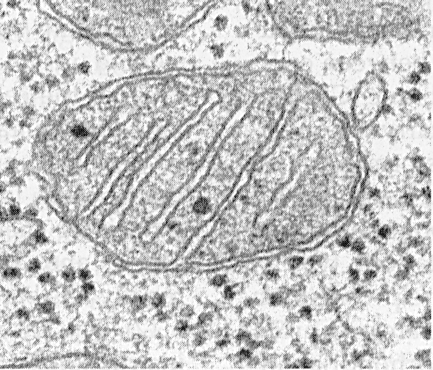
In order to diversify the festive table, a woman often tries...

Many people do not cook fish dishes because they are not very well versed in...

Homemade recipes for delicious and natural tomato sauce for barbecue...